Recovery
BPC-157: the gastric-sequence pentadecapeptide
BPC-157 (body protection compound-157) is a synthetic pentadecapeptide of 15 amino acids - sequence GEPPPGKPADDAGLV - derived from a naturally occurring protein isolated from human gastric juice. It was first characterized by researchers at the University of Zagreb, who noted that the parent protein appeared to contribute to the cytoprotective environment of the gastric mucosa. The peptide's origin in gastric juice situates it within a class of endogenous signaling molecules that the body uses to maintain mucosal integrity, a context that has guided much of the early tissue-protection research. Unlike most bioactive peptides, BPC-157 retains stability in simulated gastric conditions without requiring a carrier molecule or adjuvant, a property that has made it practical in rodent dosing studies across a range of routes including intraperitoneal injection and oral administration in drinking water. Its molecular weight is approximately 1,419 daltons. Preclinical investigation has since extended well beyond the gastrointestinal tract into musculoskeletal, vascular, and neurological tissue models.
At the molecular level, BPC-157 has been studied for its ability to upregulate vascular endothelial growth factor receptor 2 (VEGFR2) and activate the downstream VEGFR2-Akt-eNOS signaling cascade. In human umbilical vein endothelial cell (HUVEC) cultures, BPC-157 increased both mRNA and protein expression of VEGFR2 without altering VEGF-A levels, suggesting that it acts at the receptor rather than the ligand. BPC-157 was found to promote VEGFR2 internalization via dynamin-dependent endocytosis; when endocytosis was blocked pharmacologically, the pro-angiogenic effect was abolished. Time-dependent phosphorylation of Akt and eNOS followed VEGFR2 engagement. In parallel, studies of tendon fibroblasts demonstrated dose-dependent activation of focal adhesion kinase (FAK) and paxillin, cytoskeletal scaffolding proteins central to integrin-mediated cell migration. This FAK-paxillin axis appears to operate independently of direct cell proliferation, instead governing cell motility, stress survival, and F-actin reorganization.
Preclinical research in rat models has examined BPC-157 across tendon, ligament, gut, and vascular tissue. In Achilles tendon transection studies, intraperitoneal administration of BPC-157 at doses ranging from 10 picograms to 10 micrograms per kilogram body weight produced consistent improvements in biomechanical load-to-failure values, functional Achilles functional index scores, and histological markers including fibroblast density and collagen organization. Comparable results were observed in medial collateral ligament transection models across 90-day follow-up periods, with oral delivery in drinking water showing efficacy equivalent to intraperitoneal injection. In vascular ischemia models using bilateral carotid occlusion and hind limb ischemia, BPC-157 treatment was associated with accelerated restoration of blood flow as measured by laser Doppler scanning and increased vessel density on histology. Gut studies have documented resolution of mucosal lesions in various injury models, consistent with the peptide's gastric origin and cytoprotective profile. Gene expression analyses in hippocampal tissue after ischemia/reperfusion identified upregulation of Akt1, Vegfr2, Nos3, Egr1, and Src, alongside downregulation of Nos2 and Nfkb, providing a transcriptional correlate for the observed functional recovery.
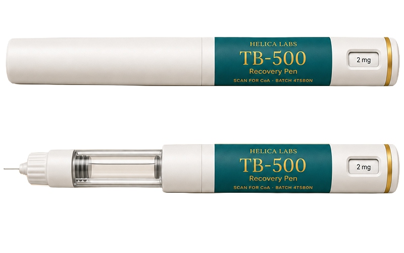
BPC-157 research continues to probe the scope of its tissue-repair activity, with studies examining bone healing, peripheral nerve regeneration, and spinal cord injury models. Its interaction with the nitric oxide system - specifically the observed shift from inducible NOS (iNOS/NOS2) toward endothelial NOS (eNOS/NOS3) gene expression - is of particular interest to researchers studying vascular remodeling, because this pattern is associated with reduced inflammatory nitric oxide production and increased vasodilatory capacity. The early growth response gene Egr-1, identified as upregulated in BPC-157-treated hippocampal tissue after ischemia, is a zinc-finger transcription factor also implicated in wound healing and angiogenesis in peripheral tissues, suggesting a conserved transcriptional response across injury types. The peptide shares partial mechanistic overlap with TB-500 (thymosin-beta-4 synthetic analog) in that both peptides influence cell motility and angiogenesis, though through distinct molecular entry points. Researchers have begun co-administering the two peptides in preclinical tissue-repair protocols to evaluate whether local angiogenesis signals from BPC-157 complement the cytoskeletal and cell-migration activities attributed to TB-500. Current literature describes BPC-157 as research-grade only; no human clinical endpoints for musculoskeletal or wound-repair indications have been established.
References:
PMID 27847966 ·
PMID 21030672 ·
PMID 14554208 ·
PMID 34267654 ·
PMID 27138887
Recovery
TB-500 and the thymosin-beta-4 actin story
TB-500 is the synthetic research analog of thymosin-beta-4 (Tbeta4), a 43-amino-acid ubiquitous peptide first isolated from calf thymus in the early 1970s and subsequently identified in virtually every nucleated mammalian cell. Thymosin-beta-4 is one of the most abundant intracellular peptides in many tissue types, with particularly high concentrations in platelets, macrophages, and wound fluid, where it is released upon cell activation or injury. Its primary structural role is the sequestration of globular actin (G-actin), which regulates the dynamic equilibrium between G-actin monomers and filamentous actin (F-actin) polymers. Because the ratio of G-actin to F-actin directly governs cytoskeletal tension and lamellipodium formation, this sequestration activity positions thymosin-beta-4 as a molecular rheostat for cell shape change and directed migration. This regulatory function places thymosin-beta-4 at the intersection of cytoskeletal organization and cell motility - processes central to wound repair, tissue remodeling, and angiogenesis. TB-500 is used in preclinical settings as a soluble, bioavailable form to study these mechanisms in cell culture and animal injury models, with the advantage that it can be administered exogenously to tissues that may have depleted endogenous thymosin-beta-4 stores after injury.
The sequestration of G-actin by thymosin-beta-4 is mediated through a central actin-binding domain containing the sequence LKKTETQ (residues 17-23). Studies using truncated and mutated peptide variants have demonstrated that this heptapeptide fragment retains the capacity to promote angiogenesis, wound healing, and epithelial cell migration, identifying it as the minimal biologically active unit within the full-length protein. Beyond simple sequestration, thymosin-beta-4 forms ternary complexes with both G-actin and profilin, influencing actin critical concentration and altering the kinetics of filament assembly at cell membranes. A landmark study published in Nature (2004) demonstrated that thymosin-beta-4 forms a functional complex with PINCH (particularly interesting new cysteine-histidine rich protein) and integrin-linked kinase (ILK), resulting in activation of the serine-threonine kinase Akt. This ILK-PINCH-Akt cascade promotes cardiomyocyte and endothelial cell survival and migration. Additionally, thymosin-beta-4 upregulates matrix metalloproteinases (MMPs), particularly MMP-1, -2, and -3, through a mechanism partially independent of its actin-binding domain; MMP activity has been shown necessary for thymosin-beta-4-stimulated epithelial cell migration.
In rodent wound healing models, thymosin-beta-4 applied topically or intraperitoneally increased wound re-epithelialization by up to 61% over saline controls at seven days post-wounding, with measurable increases in collagen deposition and angiogenesis. Wound contraction rates in treated animals exceeded controls by at least 11%. In a full-thickness skin wound model in mice, keratinocyte migration was stimulated two- to threefold in Boyden chamber assays at doses as low as 10 picograms per milliliter. A corneal wound healing model demonstrated that MMP catalytic activity is necessary for thymosin-beta-4 promotion of epithelial migration, with broad-spectrum MMP inhibitors abolishing the effect. In a dermal burn wound study, thymosin-beta-4 treatment was associated with sustained expression of heat-shock protein 70 (HSP70), which regulated F-actin remodeling in human endothelial cells and correlated with improved wound closure and vascularization. In cardiac models, coronary artery ligation in mice followed by thymosin-beta-4 treatment produced upregulation of ILK and Akt activity, enhanced early myocyte survival, and improved cardiac function at follow-up.
Research into thymosin-beta-4 has expanded to examine its role in hair follicle activation, corneal repair, and neural tissue contexts. Studies in aged rodents indicate that the angiogenic and wound-repair effects of thymosin-beta-4 are preserved even when baseline angiogenic capacity is reduced, suggesting potential relevance in tissue environments with diminished vascular response. The LKKTETQ fragment has attracted attention as a minimal pharmacophore for developing smaller synthetic variants with defined activity profiles. In the context of combined peptide research, TB-500 and BPC-157 address distinct but complementary aspects of tissue repair: TB-500 primarily governs cell motility and cytoskeletal reorganization through the ILK-Akt axis and MMP induction, whereas BPC-157 drives local angiogenesis via VEGFR2-Akt-eNOS signaling. Preclinical co-administration studies are beginning to probe whether these pathways produce additive effects on connective tissue recovery, particularly in tendon and ligament injury models.
References:
PMID 15565145 ·
PMID 20179146 ·
PMID 17348036 ·
PMID 10469335 ·
PMID 25921810
Skin
GHK-Cu: the copper peptide rebuilds the matrix
GHK-Cu is the copper(II) complex of the naturally occurring human tripeptide glycyl-L-histidyl-L-lysine (GHK). The tripeptide was first identified in human plasma in the early 1970s, where it was found to stimulate liver cell growth and protein synthesis at physiological concentrations. Subsequent work established that GHK has a binding affinity for copper(II) ions comparable to the copper-transport site on albumin, allowing it to form a stable chelate designated GHK-Cu. This complex is present endogenously in plasma, saliva, and urine, with measured plasma concentrations declining from approximately 200 nanomolar in young adults to around 80 nanomolar in older individuals - a gradient that has been linked to age-associated reductions in tissue repair capacity. GHK-Cu is classified as a matrikine: a peptide fragment that acts on extracellular matrix biology by modulating cell behavior and gene expression relevant to tissue remodeling and skin homeostasis.
At the molecular level, GHK-Cu operates through a broad network of cellular pathways. It stimulates the synthesis of collagen types I and III, elastin, and glycosaminoglycans including dermatan sulfate and chondroitin sulfate. In parallel, it modulates the activity of matrix metalloproteinases and their tissue inhibitors (TIMPs) in a manner that supports matrix remodeling rather than simple degradation - an unusual dual activity relevant to balanced tissue turnover. GHK-Cu upregulates the small leucine-rich proteoglycan decorin, a collagen fibril organizer that regulates TGF-beta signaling and is associated with reduced scar formation. Antioxidant activity has been documented through upregulation of superoxide dismutase and suppression of iron-mediated free radical generation. GHK-Cu also suppresses thromboxane formation and TGF-beta-1 secretion, reducing inflammatory signaling in the wound microenvironment. Transcriptomic analyses have identified GHK as capable of altering the expression of more than 4,000 human genes, including those regulating DNA repair, the ubiquitin-proteasome system, and anti-cancer pathways - particularly notable in COPD patient-derived cells, where GHK exposure shifted gene expression toward a repair and remodeling profile.
Preclinical research in wound healing models across rats, mice, and pigs has documented GHK-Cu acceleration of healing in skin, gastrointestinal mucosa, bone tissue, and hair follicles. In keratinocyte studies, copper-GHK increased integrin alpha-6 and beta-1 expression, promoted p63-positive basal cell populations - a marker associated with epithelial stem cell maintenance - and elevated proliferating cell nuclear antigen (PCNA) staining, consistent with enhanced regenerative activity in the basal layer. Hair follicle research has shown that GHK-Cu increases follicle size and stimulates hair growth in rodent models. In aged animal models, wound-healing improvements were observed despite reduced baseline tissue repair capacity, paralleling the pattern seen with thymosin-beta-4. In human fibroblast cultures, GHK-Cu restored replicative capacity in cells that had been exposed to radiation, suggesting a role in mitigating therapy-induced tissue damage. Controlled studies on aged skin in human subjects reported improvements in elasticity, firmness, and reduction of photodamage markers, though these observations remain at the cosmetic research level rather than the therapeutic endpoint level.
GHK-Cu research is increasingly focused on its gene-regulatory properties rather than its immediate biochemical actions. Bioinformatic analyses of GHK's effects on the human genome have positioned it as a potential modulator of aging-associated gene expression shifts - specifically, the increase in inflammatory and tissue-destructive gene activity observed with advancing age. In this framing, GHK-Cu is studied as a potential tool to investigate mechanisms by which copper-peptide complexes influence chromatin accessibility and transcription factor activity. Related research peptides in the matrikine class, such as KTTKS (a collagen-derived pentapeptide) and palmitoyl tripeptides used in dermatological research, share conceptual overlap with GHK-Cu in targeting extracellular matrix remodeling. The intersection of GHK-Cu with decorin biology and TGF-beta modulation connects it to ongoing work on fibrosis prevention and scar resolution, areas where precise control of matrix turnover is a primary research objective.
References:
PMID 26236730 ·
PMID 18644225 ·
PMID 19319546 ·
PMID 25302294
Inflammation
KPV: the alpha-MSH fragment that quiets inflammation
KPV is a tripeptide composed of lysine-proline-valine, representing the C-terminal residues 11-13 of alpha-melanocyte-stimulating hormone (alpha-MSH). Alpha-MSH is a tridecapeptide derived by post-translational processing of the proopiomelanocortin (POMC) precursor protein and is produced in keratinocytes, melanocytes, and various immune and neural cells. Its role extends well beyond melanin regulation: alpha-MSH is one of the better-characterized endogenous anti-inflammatory neuropeptides, with activity documented in models of contact dermatitis, asthma, inflammatory bowel disease, and joint inflammation. The C-terminal tripeptide KPV was identified as the minimal sequence retaining the majority of alpha-MSH's anti-inflammatory capacity. Because KPV is structurally simpler than the full tridecapeptide, it has become a preferred tool for dissecting anti-inflammatory mechanisms and for exploring peptide delivery strategies in gut and skin research models.
The mechanistic profile of KPV centers on suppression of NF-kappaB, the master transcription factor governing the expression of pro-inflammatory cytokines including TNF-alpha, IL-1beta, IL-6, and inducible nitric oxide synthase (iNOS). In keratinocyte and macrophage cultures, alpha-MSH and KPV reduce NF-kappaB activation downstream of toll-like receptor and cytokine receptor engagement, lowering the transcription of adhesion molecules and chemokine receptors. A key finding in the mechanistic literature concerns receptor independence: KPV lacks the His-Phe-Arg-Trp tetrapeptide pharmacophore required for canonical binding to melanocortin receptors (MC1R through MC5R). Despite this, KPV retains near-complete anti-inflammatory activity comparable to the full alpha-MSH sequence and displays no appreciable melanogenic action. The precise signaling pathway engaged by KPV in the absence of melanocortin receptor binding has not been fully resolved, though similarities to alpha-MSH's downstream NF-kappaB modulation suggest a receptor-independent entry point, possibly through direct intracellular peptide uptake or an as-yet uncharacterized receptor. Signaling studies in human keratinocytes have demonstrated that KPV can elicit rapid intracellular calcium responses at femtomolar to nanomolar concentrations, implicating calcium-dependent signaling cascades independent of cyclic AMP elevation.
In preclinical animal models, alpha-MSH and KPV have been studied in colitis, cutaneous inflammation, and ocular inflammation paradigms. In a dextran sulfate-induced experimental inflammatory bowel disease model in mice, alpha-MSH treatment reduced fecal blood by more than 80%, inhibited weight loss, and significantly suppressed TNF-alpha and inflammatory nitric oxide production in lower colon tissue stimulated with concanavalin A. These results established a gut anti-inflammatory profile relevant to subsequent KPV research, given that the tripeptide accounts for most of the full hormone's activity in these models. In skin models, alpha-MSH-related peptides including KPV have been shown to reduce irritant and allergic contact dermatitis, modulate inflammatory cytokine profiles in keratinocytes, and suppress leukocyte recruitment. The peptide's small size and stability compared to the full tridecapeptide make it amenable to topical and oral delivery formats, which have been explored in rodent gut inflammation studies with encouraging results on mucosal cytokine profiles.
Current research on KPV intersects with several active areas in inflammatory biology. The observation that KPV can suppress NF-kappaB without engaging conventional melanocortin receptors has motivated investigation into whether the peptide interacts with intracellular pattern-recognition pathways or inflammasome components. Related peptide K(D)PT - a derivative corresponding to IL-1beta residues 193-195 - has emerged as a structurally distinct but functionally similar tripeptide with potent anti-inflammatory effects, and comparative studies of KPV and K(D)PT are helping to map the structural requirements for receptor-independent anti-inflammatory activity. In the broader context of peptide-based anti-inflammatory research, KPV is studied alongside Thymosin-alpha-1 and LL-37, which also modulate innate immune tone through distinct receptor systems. The common thread across these peptides is the potential to selectively dampen inflammatory amplification - particularly NF-kappaB-driven cytokine cascades - without broad immunosuppression, a distinction relevant to preclinical models of chronic mucosal inflammation.
References:
PMID 15102092 ·
PMID 17934097 ·
PMID 21222263 ·
PMID 9145424
Stacks
Why researchers combine BPC-157 and TB-500
In preclinical tissue repair research, BPC-157 and TB-500 are increasingly studied together based on the premise that their mechanistic profiles address distinct but complementary phases of healing. BPC-157 (the synthetic pentadecapeptide GEPPPGKPADDAGLV) primarily acts on vascular biology: it upregulates VEGFR2 expression and internalization, activates the VEGFR2-Akt-eNOS signaling axis, and drives the formation of new capillary networks in ischemic or injured tissue. TB-500 (the synthetic thymosin-beta-4 analog) primarily governs cytoskeletal dynamics and cell motility: it sequesters G-actin through its LKKTETQ domain, activates the ILK-PINCH-Akt survival cascade, and induces matrix metalloproteinases that facilitate cell migration through fibrin and collagen matrices. The two peptides thus target different bottlenecks in the repair sequence - BPC-157 addressing vascular supply and the inflammatory-to-proliferative transition, TB-500 addressing the cell-level machinery of migration and matrix remodeling. This mechanistic complementarity has made them a common pairing in rodent musculoskeletal injury protocols.
At the molecular level, the pathways engaged by BPC-157 and TB-500 converge on Akt kinase but through different upstream signals. BPC-157 reaches Akt via receptor tyrosine kinase engagement (VEGFR2), while TB-500 reaches Akt via integrin-linked kinase (ILK) in complex with PINCH. Both result in Akt phosphorylation, which promotes cell survival, inhibits apoptotic signaling, and supports metabolic activity in stressed tissue. Downstream, BPC-157 additionally activates eNOS to generate nitric oxide, a vasodilator and angiogenic co-factor; TB-500 additionally induces MMP-1, MMP-2, and MMP-3 expression, enzymes that remodel the provisional fibrin matrix and allow advancing cell fronts to penetrate damaged tissue. Where BPC-157 also activates FAK-paxillin signaling in fibroblasts to enhance tendon cell migration, TB-500 provides a parallel actin-based motility mechanism through the LKKTETQ active fragment. Together, the two peptides appear to address both the luminal (vascular) and parenchymal (cell migration, matrix) aspects of connective tissue repair, which may explain their co-administration in musculoskeletal injury research.
Preclinical co-administration studies remain limited in number, but the rationale is supported by independent findings from each peptide's research literature. BPC-157 accelerated healing of transected rat Achilles tendon at doses ranging from 10 picograms to 10 micrograms per kilogram, with improvements in biomechanical load-to-failure, functional scoring, and collagen organization. In medial collateral ligament transection, BPC-157 was effective across intraperitoneal, oral, and topical routes at comparable dose ranges over 90 days. TB-500 (thymosin-beta-4) increased wound re-epithelialization by up to 61% in full-thickness rodent wound models, enhanced collagen deposition, and stimulated keratinocyte migration at doses starting at 10 picograms per milliliter in vitro. In burn wound models, thymosin-beta-4 sustained HSP70 expression and F-actin remodeling in endothelial cells, improving vascularization. The theoretical basis for combining these peptides rests on the argument that BPC-157 establishes the vascular infrastructure (through VEGFR2-driven angiogenesis) while TB-500 equips individual repair cells with the motility machinery (through actin dynamics and MMP induction) needed to populate the new tissue bed.
For researchers designing preclinical co-administration protocols, the published literature on each peptide individually provides a dose-response framework. BPC-157 is typically studied at microgram-to-nanogram per kilogram ranges intraperitoneally or dissolved in drinking water; TB-500 has been administered in analogous regimens at nanomolar concentrations in rodent models. Neither peptide has established pharmacokinetic interaction data in combined-dosing studies as of available published literature, and the field lacks head-to-head dose-ranging studies for the combination. The two peptides share no known structural similarity and are unlikely to compete for the same receptor or binding site, which simplifies initial combination study design. Researchers using both peptides in parallel are generally advised to reference independent dose-response data and monitor tissue-level angiogenesis markers (vessel density, VEGFR2 expression) and cell migration markers (FAK phosphorylation, MMP levels) as orthogonal readouts that reflect each peptide's distinct mechanistic contribution. No human clinical data exist for either peptide individually or in combination in musculoskeletal or wound-repair contexts; all available evidence is from cell culture or rodent injury models.
References:
PMID 27847966 ·
PMID 15565145 ·
PMID 21030672 ·
PMID 17348036 ·
PMID 34267654
GLP-1
Semaglutide and the long-acting GLP-1 agonists
Glucagon-like peptide-1 (GLP-1) is a gut-derived incretin hormone secreted by L-cells of the small intestine in response to nutrient ingestion. In healthy physiology, GLP-1 acts on pancreatic beta cells to amplify glucose-dependent insulin secretion, suppresses glucagon release from alpha cells, slows gastric emptying, and signals the hypothalamus to reduce food intake. Native GLP-1, however, is inactivated within minutes by the enzyme dipeptidyl peptidase-4 (DPP-4), making its therapeutic use impractical without chemical modification. The development of long-acting GLP-1 receptor agonists (GLP-1 RAs) has been driven by the goal of preserving these incretin actions over extended dosing intervals. Semaglutide, the most widely studied of this drug class, was engineered to address the rapid clearance of native GLP-1 through three key structural changes: an amino acid substitution at position 8 (alanine replaced by 2-aminoisobutyric acid) to resist DPP-4 degradation, an arginine-for-lysine substitution at position 34 to direct acylation to a single site, and fatty-acid lipidation at lysine-26 with a C18 dicarboxylic fatty acid linked through a gamma-glutamic acid and two aminopolyethylene glycol (OEG) spacer units.
The lipidation strategy is central to semaglutide's pharmacokinetics. The C18 fatty diacid moiety drives reversible, high-affinity binding to human serum albumin - an affinity approximately 5.6-fold greater than that of the earlier GLP-1 RA liraglutide, which uses a simpler C16 monocarboxylic fatty acid. Albumin binding protects semaglutide from renal filtration and limits access of proteases to the peptide backbone, collectively extending the plasma half-life to approximately seven days in humans following subcutaneous administration. This once-weekly pharmacokinetic profile reflects a systematic structure-activity optimization: shorter fatty acid chains (C12-C16) yielded half-lives measured in hours, while the C18 diacid with an optimized OEG linker achieved the balance of high albumin affinity, preserved GLP-1 receptor potency, and acceptable receptor off-rate needed for weekly dosing. In preclinical and early human studies, the modifications did not attenuate the ability of semaglutide to stimulate GLP-1 receptor-mediated cAMP signaling, indicating that structural half-life engineering can be achieved without major loss of pharmacodynamic activity. Postprandial studies in subjects with obesity found that once-weekly semaglutide significantly improved glucose disposal, suppressed postprandial triglycerides and VLDL by approximately 40-43%, reduced ApoB-48 excursions, and delayed first-hour gastric emptying, collectively illustrating the breadth of incretin activity retained by the lipidated molecule.
The SUSTAIN clinical trial program evaluated subcutaneous semaglutide (0.5 mg and 1.0 mg weekly) across a spectrum of type 2 diabetes populations and comparator regimens. Across SUSTAIN 1 through 7, semaglutide consistently produced superior reductions in HbA1c compared with placebo and active comparators including sitagliptin, exenatide extended-release, and insulin glargine, with reductions ranging from approximately 1.0% to 1.8% from baseline. In SUSTAIN 6, which enrolled patients at high cardiovascular risk, once-weekly semaglutide reduced the composite endpoint of major adverse cardiovascular events by 26% relative to placebo (hazard ratio 0.74), establishing cardiovascular non-inferiority and suggesting a cardioprotective signal. The STEP clinical trial program subsequently evaluated semaglutide at the higher dose of 2.4 mg per week in persons with obesity without diabetes. In STEP 1, participants treated with semaglutide 2.4 mg plus lifestyle intervention achieved a mean body weight reduction of 14.9% at 68 weeks, compared with 2.4% in the placebo group, with 86.4% of the semaglutide group achieving at least 5% weight loss. These data established subcutaneous semaglutide 2.4 mg as a benchmark for pharmacological weight reduction in the GLP-1 RA class.
Semaglutide exists in both subcutaneous and oral formulations, the latter co-formulated with the absorption enhancer sodium N-(8-[2-hydroxybenzoyl] amino)caprylate (SNAC) to permit gastric absorption. The oral form demonstrated a similar half-life of approximately one week and supported the PIONEER trial program showing meaningful HbA1c reductions in type 2 diabetes. The development of semaglutide has informed the design of subsequent long-acting GLP-1 RAs and multi-receptor agonists, with the C18 fatty diacid lipidation motif serving as a prototype for acylation strategies applied to tirzepatide and related molecules. Research into depot formulations, including hydrogel microsphere encapsulation, is exploring whether semaglutide's half-life could be further extended to once-monthly dosing with preserved lean-mass-sparing weight reduction. The broader GLP-1 RA class - encompassing liraglutide, dulaglutide, exenatide LAR, and albiglutide - shares the incretin biology of semaglutide but differs in half-life engineering strategies, and semaglutide's pharmacokinetic and clinical profile has made it the current reference comparator for emerging incretin-based agents.
References:
PMID 28941314 ·
PMID 33567185 ·
PMID 30615985
GLP-1
Tirzepatide: the first dual GIP/GLP-1 agonist
Tirzepatide (LY3298176) is a synthetic acylated peptide that co-activates two incretin receptors: the glucose-dependent insulinotropic polypeptide receptor (GIPR) and the glucagon-like peptide-1 receptor (GLP-1R). It was engineered by grafting GLP-1 receptor agonist activity into the native GIP sequence, producing a 39-amino-acid peptide attached to a C20 fatty diacid chain via a gamma-glutamic acid and two mini-PEG spacers at a lysine residue - a lipidation strategy that confers a plasma half-life of approximately five days in humans and supports once-weekly subcutaneous dosing. GIP, secreted from K-cells of the proximal small intestine, is one of the two major incretin hormones alongside GLP-1. While GIP is a potent stimulator of glucose-dependent insulin secretion, its actions extend to adipose tissue - where GIPR activation promotes lipolysis and lipid storage modulation - and to central nervous system circuits involved in energy balance. Co-engagement of both incretin receptor systems in a single molecule was hypothesized to amplify metabolic benefits beyond those achievable through GLP-1R agonism alone, motivating tirzepatide's development as the first approved dual incretin receptor co-agonist.
Pharmacological characterization of tirzepatide at the molecular level reveals an asymmetric receptor engagement profile. At the GIPR, tirzepatide displays affinity and cAMP-generating potency equivalent to native GIP, with full agonist activity and comparable receptor internalization kinetics. At the GLP-1R, tirzepatide exhibits approximately 5-fold lower binding affinity and 20-fold lower cAMP potency than native GLP-1, functioning as a partial agonist in low-receptor-density assays. Notably, tirzepatide shows biased signaling at the GLP-1R, strongly favoring cAMP accumulation while demonstrating less than 10% efficacy for beta-arrestin2 recruitment and markedly reduced GLP-1R internalization compared with native GLP-1 or selective GLP-1R agonists. This biased agonism is pharmacologically significant because beta-arrestin1 limits the insulin secretory response to GLP-1 in pancreatic islets; by bypassing arrestin recruitment at the GLP-1R, tirzepatide may preserve a more sustained insulinotropic signal. In human islets, studies demonstrated that blocking either receptor with selective antagonists partially attenuates tirzepatide-stimulated insulin secretion, confirming that the dual-receptor mechanism contributes to hormone secretion from human beta cells. Receptor occupancy modeling using clinically relevant dosing indicated that tirzepatide engages the GIPR more fully than the GLP-1R at steady-state concentrations, underpinning its characterization as an imbalanced, biased dual agonist rather than a simple equipotent co-stimulator.
The SURPASS clinical trial program evaluated tirzepatide (5 mg, 10 mg, and 15 mg weekly) in five phase 3 trials in persons with type 2 diabetes using a range of comparators. Across SURPASS 1 through 5, tirzepatide reduced HbA1c from baseline by 1.87% to 2.07% (SURPASS-1, placebo comparison) and demonstrated superiority over semaglutide 1.0 mg in direct comparison, with HbA1c reductions up to 2.58% and body weight reductions of 5.4 to 11.7 kg across the program. A sizable proportion of SURPASS participants - 23% to 62% - achieved HbA1c values below 5.7%, the upper limit of normoglycemia. In the SURMOUNT-1 trial, tirzepatide was evaluated for weight management in adults with obesity or overweight plus a comorbidity, without diabetes. At 72 weeks, tirzepatide 10 mg and 15 mg produced mean weight reductions of 19.5% and 20.9%, respectively, versus 3.1% with placebo; 57% of participants on the 15 mg dose achieved at least 20% body weight reduction. Tirzepatide also improved insulin sensitivity - as assessed by homeostasis model assessment of insulin resistance (HOMA-IR) - beyond what was explained by weight loss alone, consistent with the hypothesis that GIP receptor agonism in adipose tissue contributes independent metabolic effects.
The question of whether tirzepatide's clinical advantages over selective GLP-1R agonists reflect additive or synergistic incretin signaling remains an active area of research. Preclinical studies in rodent islets showed that, when GLP-1R was genetically absent, tirzepatide's insulinotropic response was fully blocked by a GIPR antagonist, and vice versa, indicating that both receptor pathways are independently necessary and functionally cooperative. Whether the combined stimulation produces effects that exceed what would be predicted by dose-matched monotherapies - i.e., true synergy - is difficult to disentangle from the pharmacokinetic and receptor-density complexities of the dual-agonist system. From a clinical standpoint, tirzepatide consistently outperformed semaglutide in head-to-head comparisons, though differences in dose-matched receptor engagement make mechanistic attribution challenging. Subsequent research is examining tirzepatide's effects on liver steatosis, sleep apnea, heart failure, and chronic kidney disease, and the molecule has served as the structural template informing the design of triple receptor agonists such as retatrutide that add glucagon receptor activity to the GIP and GLP-1 axes.
References:
PMID 32730231 ·
PMID 34186022 ·
PMID 35658024 ·
PMID 37277609
GLP-1
Retatrutide and triple agonism (GLP-1 + GIP + glucagon)
Retatrutide (LY3437943) is a synthetic acylated peptide that activates three metabolically relevant receptors simultaneously: the glucagon-like peptide-1 receptor (GLP-1R), the glucose-dependent insulinotropic polypeptide receptor (GIPR), and the glucagon receptor (GCGR). It represents the first triple incretin receptor agonist to enter and complete phase 2 clinical evaluation. The molecule was designed on the recognition that dual GIP/GLP-1 co-agonism, as demonstrated by tirzepatide, produces weight loss and glycemic improvements beyond those achieved by selective GLP-1R agonism, and that adding GCGR activity could further amplify energy expenditure - a mechanism not meaningfully engaged by GIP or GLP-1 receptor stimulation alone. Glucagon, primarily known as a counter-regulatory hormone that raises blood glucose by stimulating hepatic glycogenolysis and gluconeogenesis, also activates brown adipose tissue thermogenesis and increases resting energy expenditure when administered acutely. The theoretical challenge of adding GCGR agonism to a molecule that also activates GLP-1R and GIPR is that the hyperglycemic action of glucagon must be counterbalanced by the insulinotropic and glucagon-suppressing actions of GLP-1 and GIP, such that net glucose homeostasis remains adequate. Retatrutide's design embodies this pharmacological balancing act, with the GLP-1R and GIPR components providing glycemic counterregulation to the GCGR-mediated metabolic stimulus.
The GCGR component of retatrutide's activity recruits distinct mechanisms from those engaged by GLP-1 or GIP receptor activation. GCGR stimulation in the liver and adipose tissue increases cAMP, activating hormone-sensitive lipase and promoting triglyceride breakdown. In brown and beige adipose tissue, GCGR agonism increases expression of uncoupling protein 1 (UCP-1) and thermogenic gene programs, translating into elevated resting energy expenditure. This thermogenic contribution is a distinguishing feature of triple receptor agonism: whereas tirzepatide's weight loss is primarily driven by reduced energy intake through combined hypothalamic GIP and GLP-1 receptor signaling, retatrutide is proposed to additionally increase the energy expenditure side of the energy balance equation through GCGR activity. In obese rodent models and early mechanistic studies, glucagon receptor agonism delayed gastric emptying synergistically with GLP-1R activation, reduced food intake, and lowered body fat depots including visceral adiposity. The C18 fatty diacid lipidation of retatrutide, attached through a linker to a lysine residue, confers a pharmacokinetic profile suitable for once-weekly subcutaneous dosing, consistent with the lipidation-based half-life strategies used for semaglutide and tirzepatide.
Phase 2 clinical data for retatrutide have been published from two trials. In a 48-week randomized, placebo-controlled phase 2 trial in adults with obesity (without diabetes), retatrutide at 12 mg weekly produced a mean body weight reduction of 24.2% - a magnitude of weight loss approaching that seen with some bariatric surgical procedures and representing the largest pharmacologically achieved weight reduction reported in a randomized trial at the time of publication. Dose-dependent reductions were observed across all active retatrutide groups (1 mg, 4 mg, 8 mg, and 12 mg), with 100% of participants in the 8 mg and 12 mg groups achieving at least 5% body weight reduction at 48 weeks. In a separate phase 2 trial in adults with type 2 diabetes, retatrutide reduced HbA1c by up to 2.02% at 24 weeks and body weight by up to 16.94% at 36 weeks compared with baseline, significantly exceeding both placebo and the active comparator dulaglutide 1.5 mg. A meta-analysis of available randomized controlled trial data showed retatrutide significantly reduced body weight by a weighted mean of 10.66 kg compared with placebo, alongside reductions in BMI and waist circumference. Gastrointestinal adverse events (nausea, vomiting, diarrhea, constipation) were dose-dependent, mild to moderate in severity, and consistent in character with the GLP-1 RA class; no severe hypoglycemia or deaths were reported in phase 2 studies.
The clinical weight loss signal from retatrutide has reframed expectations for pharmacological obesity treatment and accelerated phase 3 investigation. The TRANSCEND and TRIUMPH trial programs are evaluating retatrutide in type 2 diabetes and obesity, respectively, at scale and over longer durations. A key mechanistic question under active investigation is the quantitative contribution of the GCGR component to weight loss beyond that attributable to dual GIP/GLP-1R activation: since tirzepatide already achieves approximately 20-21% weight loss at maximal doses, the incremental weight loss with retatrutide implies that the glucagon receptor arm adds clinically meaningful energy expenditure. Body composition analyses from the type 2 diabetes phase 2 trial showed that the ratio of lean to fat mass loss with retatrutide was similar to that observed with other approved obesity pharmacotherapies, suggesting that the additional weight loss from glucagon receptor agonism came disproportionately from fat mass rather than lean tissue - an important safety consideration for skeletal muscle preservation. Retatrutide's development has established triple receptor agonism as a viable drug class and has prompted exploration of next-generation molecules targeting the GLP-1R alongside receptors for amylin, glucagon, and other metabolic peptides.
References:
PMID 37366315 ·
PMID 37385280 ·
PMID 39318607
Metabolic
Cagrilintide and the amylin axis
Amylin (islet amyloid polypeptide, IAPP) is a 37-amino-acid peptide hormone co-secreted with insulin from pancreatic beta cells in response to meals. Its physiological roles in the regulation of energy balance are distinct from those of insulin, GLP-1, or GIP: amylin acts primarily through amylin receptors (AMY receptors, which are heterodimers of the calcitonin receptor and receptor activity-modifying proteins RAMP1, RAMP2, or RAMP3) in the area postrema of the caudal hindbrain, a circumventricular organ that lacks a blood-brain barrier and is therefore directly accessible to circulating peptides. From the area postrema, amylin signal propagates through the nucleus of the solitary tract and lateral parabrachial nucleus, converging on hypothalamic circuits that regulate meal size, gastric emptying rate, and overall energy intake. In persons with type 2 diabetes, beta-cell amylin secretion is impaired, paralleling the loss of insulin secretory capacity, and in type 1 diabetes it is virtually absent. Native human amylin is prone to aggregation and fibrillization, which limited its direct therapeutic development and necessitated the synthesis of non-aggregating analogs. Pramlintide, based on the non-fibrillating rat amylin sequence, was the first clinically approved amylin analog, used as an adjunct to insulin therapy; its short half-life requiring multiple daily injections constrained its broader application.
Cagrilintide is a long-acting, non-fibrillating synthetic amylin analog engineered for once-weekly subcutaneous administration. Its design incorporates fatty acid acylation - analogous to the lipidation strategy used for semaglutide - to enable albumin binding and extended plasma half-life, alongside backbone modifications that prevent the self-assembly into amyloid-like fibers that occurs with native IAPP. Cagrilintide binds AMY1R and AMY3R receptor subtypes in the area postrema and hindbrain, producing sustained anorectic signaling without the acute receptor desensitization seen with short-acting amylin analogs. In preclinical studies using wild-type mice and receptor-knockout models, cagrilintide produced significant dose-dependent reductions in food intake and body weight, with the weight loss concentrated in fat mass while lean mass was largely preserved. When RAMP1/3 knockout mice - which lack functional AMY1R and AMY3R - were treated with cagrilintide, the anorectic and weight-reducing effects were abolished, demonstrating that the satiety signaling depends specifically on canonical amylin receptor subtypes rather than off-target calcitonin receptor activation. This mechanistic selectivity is important because it positions cagrilintide's satiety signals as neurally distinct from, and potentially complementary to, those of GLP-1 receptor agonists, which act primarily through GLP-1Rs in the hypothalamic arcuate nucleus and vagal afferents.
The phase 2 dose-finding trial of cagrilintide monotherapy (CALM trial) enrolled adults with obesity or overweight plus comorbidity, randomized to once-weekly cagrilintide at doses from 0.3 to 4.5 mg or comparators. At 26 weeks, cagrilintide produced dose-dependent body weight reductions of 6.0% to 10.8% versus 3.0% with placebo. The highest cagrilintide dose (4.5 mg) produced weight loss comparable to liraglutide 3.0 mg daily, with a generally manageable tolerability profile dominated by gastrointestinal adverse events including nausea and constipation. The combination of cagrilintide with semaglutide - designated CagriSema - was evaluated based on the hypothesis that engaging two mechanistically independent satiety pathways (amylin receptor and GLP-1R) would produce additive or more-than-additive weight reduction. Clinical data from phase 2 CagriSema trials showed that the fixed-dose combination (cagrilintide 2.4 mg plus semaglutide 2.4 mg once weekly) produced significantly greater percentage weight loss than semaglutide 2.4 mg alone, with mean differences in weight reduction of approximately 7-9 percentage points across trials. A phase 3 trial (REDEFINE 1) subsequently demonstrated that CagriSema produced mean weight loss of approximately 22.7% versus 7.5% with placebo at 68 weeks, with 29% of CagriSema participants achieving at least 30% body weight reduction. These findings corroborate the additive satiety hypothesis and have driven interest in multi-axis appetite regulation as a strategy for obesity pharmacotherapy.
Amylin analogs beyond cagrilintide currently under investigation include petrelintide, eloralintide, and Met-233, each representing distinct structural approaches to long-acting IAPP analog design. A conceptually further-reaching development is amycretin, a unimolecular amylin receptor and GLP-1R co-agonist combining both pharmacologies in a single peptide backbone - an approach analogous to how tirzepatide unified GIP and GLP-1 agonism. Beyond weight management, the amylin axis is studied for its potential to modulate prandial glucagon secretion, reduce postprandial blood glucose excursions, and contribute to the management of non-alcoholic steatohepatitis, given beta-cell amylin deficiency in metabolic liver disease contexts. Research continues to characterize the interaction between AMY receptor subtypes (AMY1R, AMY2R, AMY3R) and downstream neuronal circuits, and to define how amylin receptor agonism integrates with the broader neuroendocrine network that includes leptin, insulin, and GLP-1 receptor signaling in the regulation of long-term body weight.
References:
PMID 34798060 ·
PMID 36883831 ·
PMID 39317404
Metabolic
AOD-9604: the hGH fragment 176-191 lipolysis story
Human growth hormone (hGH) is a 191-amino-acid anterior pituitary peptide with broad metabolic effects, including promotion of lipolysis, inhibition of lipogenesis, and modulation of adipose tissue substrate utilization. These metabolic effects have long been recognized as distinct from hGH's growth-promoting actions, which are mediated through IGF-1 production following hGH receptor activation. Research into hGH's structure-activity relationships identified the C-terminal region - roughly encompassing residues 176 through 191 - as a domain with intrinsic lipolytic activity that could be separated from the mitogenic, diabetogenic, and IGF-1-stimulating properties of the intact hormone. AOD-9604 is a synthetic peptide analog of this C-terminal fragment, corresponding to hGH residues 176 to 191 with a tyrosine-to-phenylalanine substitution at the C-terminus and a tyrosine residue added at the N-terminus to enhance stability. An earlier related compound, AOD-9401 (sometimes referred to as hGH fragment 177-191), was used in foundational metabolic studies; AOD-9604 represents the refined, more stable version of this lipolytic hGH sub-sequence. Preclinical research has studied AOD-9604 for its ability to stimulate fat breakdown and reduce fat accumulation in animal models without engaging the hGH receptor, and therefore without producing the insulin resistance, glucose intolerance, or cell proliferative effects associated with intact hGH administration.
A central finding from preclinical studies of AOD-9604 is that it does not bind the classical hGH receptor and does not stimulate cell proliferation through hGH receptor-mediated pathways, yet retains the lipolytic and antilipogenic activity of the intact hormone. In vitro receptor binding assays using cells transfected with the hGH receptor confirmed that AOD9604 fails to compete with 125I-labeled hGH for receptor occupancy, and AOD9604 did not induce the cell proliferation characteristic of hGH receptor activation. The lipolytic mechanism of AOD-9604 therefore involves a receptor or signaling pathway distinct from the canonical hGH receptor. Investigations into beta-adrenergic system involvement revealed that chronic treatment with both hGH and AOD9604 in obese mice upregulated expression of beta-3 adrenergic receptor (beta-3-AR) messenger RNA in adipose tissue. The beta-3 adrenergic receptor is the primary lipolytic receptor in rodent adipocytes and an important regulator of thermogenesis in brown adipose tissue. To determine whether beta-3-AR upregulation was mechanistically necessary for AOD9604's lipolytic effects, studies were conducted in beta-3-AR knockout mice: in these animals, the chronic reductions in body weight and lipolysis seen in wild-type mice treated with hGH or AOD9604 were absent, indicating that beta-3-AR expression is required for the long-term lipolytic response. Importantly, in an acute experiment, AOD9604 retained the ability to increase energy expenditure and fat oxidation even in beta-3-AR knockout mice, suggesting that the acute metabolic action involves a beta-3-AR-independent pathway, while the chronic weight-reducing effect depends on sustained upregulation of beta-3-AR in adipose tissue.
Systematic preclinical studies examined the metabolic effects of AOD-9604 and the closely related AOD-9401 across multiple rodent models of obesity. In obese ob/ob mice and Zucker fatty rats, oral and intraperitoneal administration of AOD-9604 or AOD-9401 produced significant reductions in body weight gain without altering food consumption, indicating that the effect was mediated through changes in energy expenditure and substrate oxidation rather than appetite suppression. Indirect calorimetry in ob/ob mice showed acute increases in energy expenditure and fat oxidation rates following treatment with AOD-9604, accompanied by elevated plasma glycerol levels - a direct index of lipolysis in adipose tissue. In adipose tissue assays conducted ex vivo, treatment increased lipolytic activity and reduced lipogenic activity, consistent with a shift in adipose tissue metabolism toward net fat mobilization. Crucially, unlike intact hGH, AOD-9604 and AOD-9401 did not induce hyperglycemia, did not reduce insulin secretion, and did not impair euglycemic clamp-assessed insulin sensitivity in treated animals. In Zucker rats treated with oral AOD-9604 at 500 micrograms per kilogram body weight daily for 19 days, body weight gain was reduced by more than 50% compared with controls, again without glucose tolerance impairment. Studies using isolated human adipose tissue demonstrated that these lipolytic and antilipogenic effects translate to human tissue in vitro, finding that the peptide increased lipolytic activity and decreased lipogenesis in explanted adipocytes from obese human donors.
The mechanistic pathway through which AOD-9604 signals to produce lipolysis - independent of the hGH receptor and at least partially independent of acute beta-3-AR activation - remains an area of ongoing inquiry. Some data suggest interaction with a beta-3-AR-related or novel receptor on adipocytes that is not the classical hGH receptor, and that the peptide may function partly through modulation of hormone-sensitive lipase and perilipin regulatory networks within the adipocyte. The preclinical lipolysis story established AOD-9604 as a candidate for investigating selective fat-metabolism modulation without the hyperglycemic and proliferative liabilities of full-length hGH. Related peptides under research investigation include other hGH C-terminal fragments and modified AOD sequences designed to optimize stability and receptor-level selectivity. The broader context of hGH fragment research has expanded to examine whether discrete domains of the hGH sequence regulate different aspects of metabolism - including cartilage repair and anabolic tissue responses - lending the hGH fragment field relevance beyond lipolysis. AOD-9604 remains studied as a biochemical probe for understanding the relationship between growth hormone biology, adipose tissue lipolytic machinery, and the beta-adrenergic regulatory network that controls fat mobilization and thermogenic capacity.
References:
PMID 11146367 ·
PMID 11713213 ·
PMID 11673763 ·
PMID 10950816
GH
GHRH analogs: Sermorelin, CJC-1295, Tesamorelin compared
Growth hormone-releasing hormone (GHRH) is a 44-amino-acid hypothalamic neuropeptide that stimulates somatotroph cells in the anterior pituitary to synthesize and secrete growth hormone. Three synthetic analogs have been studied extensively: Sermorelin, a truncated 29-amino-acid form comprising the biologically active N-terminal sequence of endogenous GHRH; CJC-1295, a tetrasubstituted derivative of GHRH(1-29) engineered to bind covalently to circulating serum albumin; and Tesamorelin, a full-length 44-residue form of GHRH modified at its N-terminus with a trans-3-hexenoic acid moiety to resist enzymatic cleavage. All three bind the GHRH receptor (GHRHR), a Gs-coupled seven-transmembrane receptor expressed on somatotrophs, activating adenylate cyclase, elevating intracellular cyclic AMP, and triggering calcium-dependent GH exocytosis. Each analog preserves the feedback architecture of the GH axis - somatostatin continues to modulate pituitary output, meaning GH secretion remains subject to negative feedback in a way that exogenous recombinant GH administration does not permit.
The core mechanistic distinction among the three analogs is their half-life, which determines the pulsatility profile of GH secretion they can produce. Sermorelin has a plasma half-life of roughly 10-20 minutes, closely approximating the rapid clearance of native GHRH and favoring pulsatile administration. A D-alanine substitution at position 2 of GHRH(1-29), studied by Soule and colleagues, reduces metabolic clearance by approximately 47% and extends the disappearance half-time from around 4.3 to 6.7 minutes in human subjects, illustrating how even single residue modifications affect pharmacokinetics. Tesamorelin achieves extended bioactivity through its hexenoyl N-terminal modification, which renders the molecule resistant to cleavage by dipeptidyl aminopeptidase IV, an enzyme that rapidly inactivates unmodified GHRH. CJC-1295 takes a fundamentally different approach: a maleimido linker at its C-terminus reacts in vivo with the free thiol on cysteine-34 of serum albumin, forming a stable bioconjugate with a measured plasma half-life of 5.8 to 8.1 days in healthy human subjects. This drug-affinity complex (DAC) technology means a single subcutaneous dose of CJC-1295 produces dose-dependent increases in mean GH concentrations for six or more days and elevates IGF-1 for nine to eleven days, findings documented in a randomized, placebo-controlled, ascending-dose trial.
Preclinical and early clinical studies have characterized each analog's downstream effects. CJC-1295 has been shown in a pharmacodynamic study to elevate trough and mean GH levels while preserving the natural pulsatile frequency of GH secretory bursts - an important distinction because pulse-dependent STAT5b signaling in liver and peripheral tissues is required for full IGF-1 production and anabolic signaling. After multiple weekly doses of CJC-1295, mean IGF-1 levels remained elevated for up to 28 days. Tesamorelin's clinical development focused on HIV-associated lipodystrophy, a condition characterized by visceral fat accumulation linked to relative GH deficiency in people receiving antiretroviral therapy. In a 12-month randomized placebo-controlled trial in 404 HIV-infected patients, tesamorelin at 2 mg/day reduced visceral adipose tissue by approximately 18% at 52 weeks, improved triglyceride profiles, and produced no significant perturbation of fasting glucose, consistent with stimulation of endogenous pulsatile GH rather than supraphysiological continuous exposure. Sermorelin research has focused on pituitary responsiveness in growth hormone-deficient states, where its short half-life necessitates frequent dosing but allows close approximation of physiological nocturnal GH pulses.
Tesamorelin is the only GHRH analog to receive FDA approval, indicated specifically for the reduction of excess abdominal fat in adults with HIV and lipodystrophy (approved November 2010, brand name Egrifta). Sermorelin has FDA-approved use as a diagnostic agent for GH reserve assessment. CJC-1295 has not proceeded beyond early-phase human pharmacokinetic studies and remains a research compound. Across all three, current research questions center on the long-term consequences of chronically elevated IGF-1, given the established relationships between IGF-1 signaling and tissue proliferation. The mechanistic differences among these analogs - half-life, receptor occupancy pattern, and downstream IGF-1 kinetics - continue to inform the design of next-generation GHRH receptor agonists aimed at more selective modulation of the somatotropic axis.
References:
PMID 15817669 ·
PMID 7962295 ·
PMID 16352683 ·
PMID 7892191 ·
PMID 20101189
GH
GHRPs and the ghrelin receptor: Ipamorelin, GHRP-2, GHRP-6, Hexarelin
Growth hormone-releasing peptides (GHRPs) are a class of synthetic, non-natural peptides that stimulate GH release through a receptor distinct from the GHRH receptor. Their endogenous cognate is ghrelin, a 28-amino-acid acylated peptide produced primarily by the stomach, and the receptor they share - designated GHS-R1a (growth hormone secretagogue receptor type 1a) - is a constitutively active, Gq/11-coupled G-protein receptor expressed on somatotrophs and in the hypothalamus. Four GHRPs have been studied in detail: GHRP-6 (His-D-Trp-Ala-Trp-D-Phe-Lys-NH2), the original reference hexapeptide; GHRP-2 (D-Ala-D-beta-Nal-Ala-Trp-D-Phe-Lys-NH2), a non-natural super-analog with markedly greater potency; Hexarelin (His-D-2-methylTrp-Ala-Trp-D-Phe-Lys-NH2), a hexapeptide analog with additional N-methyl modification conferring superior GH-releasing efficacy in vivo; and Ipamorelin (Aib-His-D-2-Nal-D-Phe-Lys-NH2), a pentapeptide identified through systematic structure-activity work that intentionally lacks the central Ala-Trp dipeptide of GHRP-1.
At the receptor level, GHRPs function as orthosteric agonists at GHS-R1a, competing with ghrelin for the same binding pocket rather than acting as allosteric modulators, as was initially proposed for some secretagogues. GHS-R1a activation stimulates phospholipase C through Gq/11, increasing inositol triphosphate and diacylglycerol, mobilizing intracellular calcium, and triggering GH vesicle fusion. GHRPs also act at the hypothalamus to amplify GHRH outflow and partially antagonize somatostatin inhibition, producing synergistic effects when co-administered with GHRH - a relationship that has been exploited in clinical testing of combined GHRH-GHRP protocols. The selectivity profiles of the four peptides differ substantially with respect to co-secretion of other pituitary hormones. GHRP-2 and Hexarelin both produce dose-dependent increases in GH but also stimulate prolactin, ACTH, and cortisol release. In a direct comparison in healthy adults, intravenous GHRP-2 and Hexarelin at 1 and 2 micrograms/kg elicited GH responses exceeding those of GHRH itself, while simultaneously inducing prolactin elevations comparable to thyrotropin-releasing hormone and ACTH/cortisol rises similar in magnitude to corticotropin-releasing hormone infusion.
Hexarelin's age-dependent GH response was characterized systematically across prepubertal children, pubertal adolescents, young adults, and elderly subjects: peak GH secretion occurred in pubertal children (mean AUC 1960 micrograms·min/l) and young adults, with blunted responses in both prepubertal children and the elderly - a finding consistent with puberty-related changes in somatostatin tone and pituitary somatotroph sensitivity. Hexarelin's prolactin- and ACTH-stimulating properties were relatively stable across age groups. Ipamorelin's selectivity profile stands in contrast: at doses more than 200-fold above its GH-release ED50, Ipamorelin produced no statistically significant elevation of ACTH, cortisol, prolactin, FSH, LH, or TSH in rat and swine models. This selectivity, comparable to GHRH itself, distinguished Ipamorelin from all earlier GHRPs and was attributed to structural features that reduce binding to non-GHS-R1a targets involved in corticotroph and lactotroph signaling. GHRP-6, the reference compound, occupies an intermediate position: it stimulates GH potently but causes measurable cortisol and prolactin co-secretion at clinical doses.
The selectivity differences among GHRPs have directed research toward Ipamorelin as the preferred candidate for long-duration studies where off-target hormone elevations could confound metabolic or sleep endpoints. GHRP-2, by contrast, has been used extensively as a pharmacological probe for pulsatile GH dynamics in neuroendocrine research because its robust, non-selective stimulation allows near-maximal GH secretion when paired with GHRH or arginine, and simultaneous GHRH-GHRP-2 infusion produces synergistic pulsatile GH output that exceeds additive expectations. Hexarelin's sleep studies demonstrated that repeated nocturnal dosing at four 50-microgram doses between 22:00 and 01:00 reduced stage-4 slow-wave sleep and EEG delta power during the total night, increased GH and prolactin concentrations across the night, and raised ACTH and cortisol during the first half of the night - illustrating the physiological cost of its broad receptor engagement compared to the more selective GHRPs. GHRP-6, the original reference hexapeptide, remains important in receptor pharmacology studies: binding competition assays using radiolabeled ghrelin have confirmed that GHRP-6 and other GHRPs occupy the orthosteric site, fully displacing labeled ghrelin at saturating concentrations and providing no evidence for allosteric modulation of ghrelin's binding kinetics. Current preclinical research is exploring GHS-R1a biology beyond GH secretion, including roles in energy homeostasis, gastrointestinal motility, neuroprotection, and hippocampal memory consolidation, all of which are functions attributable to the endogenous ghrelin/GHS-R1a system that these peptides pharmacologically engage. The structural diversity of the GHRP class - from the hexapeptide GHRP-6 and Hexarelin to the pentapeptide Ipamorelin - also provides a scaffold for understanding which molecular features drive selectivity at GHS-R1a versus related receptors, an area of ongoing medicinal chemistry investigation.
References:
PMID 9849822 ·
PMID 9285939 ·
PMID 9437229 ·
PMID 15177700
GH
Why CJC-1295 + Ipamorelin is the most-studied pulse-style stack
The rationale for combining a GHRH analog with a ghrelin receptor agonist is rooted in the distinct receptor systems and cellular mechanisms through which they each amplify GH secretion. GHRH analogs such as CJC-1295 act on pituitary somatotrophs via the GHRH receptor (GHRHR), a Gs-coupled receptor linked to cyclic AMP accumulation and protein kinase A activation. GHRPs such as Ipamorelin act on GHS-R1a, a Gq/11-linked receptor whose activation increases intracellular calcium through phospholipase C. Because these pathways converge on GH exocytosis through independent intracellular second messengers, co-stimulation produces synergistic rather than merely additive GH release - a pharmacological principle demonstrated in multiple preclinical and clinical protocols involving simultaneous infusion of GHRH and GHRP-2. The pairing of CJC-1295 with Ipamorelin specifically has attracted research interest because CJC-1295 provides prolonged, stable elevation of GHRH receptor occupancy through its albumin-binding (DAC) modification, while Ipamorelin adds pulsatile GHS-R1a activation with high GH selectivity and without the prolactin or cortisol co-secretion seen with other GHRPs.
The pulsatile versus tonic distinction is mechanistically important. Endogenous GH is secreted in discrete bursts, primarily during early slow-wave sleep, with intervening troughs near or at the assay detection limit. This pulsatility drives the pulsed activation of STAT5b in the liver and peripheral tissues, which is essential for sex-differentiated IGF-1 production patterns. Continuous high GH exposure, as occurs with pharmacological recombinant GH, blunts STAT5b signaling through receptor desensitization. A key human pharmacokinetic study of CJC-1295 demonstrated that despite the compound's extended albumin-bound half-life of 5.8 to 8.1 days, single-dose administration preserved the pulsatile frequency and amplitude of GH secretory bursts while markedly elevating trough GH levels - the basal interburst GH concentration increased 7.5-fold compared to pre-treatment - and raising mean IGF-1 by approximately 45%. When Ipamorelin is added to this background of elevated GHRH tone, each Ipamorelin pulse is expected to produce amplified GH secretion because the pituitary somatotroph pool is already primed by sustained GHRH signaling.
Preclinical pairing logic draws on the well-characterized synergy between the GHRH and GHRP systems. Studies using simultaneous intravenous infusion of GHRH and GHRP-2 in healthy older men have shown that GHRP-2 has an effect size of approximately 89 to 105 micrograms per liter per 10 hours on pulsatile GH output above the GHRH-alone condition, and that this synergistic increment was independent of testosterone and estradiol status. IGF-1 elevation following CJC-1295 alone was correlated with proteomic changes in serum consistent with activation of GH-dependent hepatic signaling, including changes in apolipoprotein A1 and transthyretin isoforms identifiable by two-dimensional gel electrophoresis. Adding a selective GHRP to the CJC-1295 background further amplifies the acute GH pulse without adding corticotroph or lactotroph stimulation, which is the pharmacological basis for the selectivity advantage of Ipamorelin over GHRP-2 or Hexarelin in long-term stacking protocols. Ipamorelin's EC50 for GH release in rat pituitary cells is approximately 1.3 nanomoles per liter, with efficacy comparable to GHRP-6, and it fails to stimulate ACTH or cortisol at doses exceeding 200-fold its GH-effective dose.
The downstream IGF-1 axis provides the principal biomarker monitored in CJC-1295 and Ipamorelin research. IGF-1 produced primarily in the liver under GH drive mediates anabolic effects in skeletal muscle, bone, and adipose tissue, and its circulating level reflects cumulative GH exposure integrated over days to weeks. After multiple weekly doses of CJC-1295, mean IGF-1 remained above pre-treatment baseline for up to 28 days, indicating that even infrequent dosing of the DAC-modified analog sustains meaningful somatotropic axis stimulation. Current research directions include understanding how the ratio of baseline-to-peak GH exposure - shaped by CJC-1295's elevation of trough levels and Ipamorelin's bolus pulse amplification - translates into specific tissue-level responses. Open questions include whether preserving pulsatility with this combination avoids the hepatic insulin resistance associated with continuous exogenous GH, and what the long-term IGF-1 implications are for individuals with pre-existing metabolic abnormalities.
References:
PMID 16352683 ·
PMID 17018654 ·
PMID 19386527 ·
PMID 9849822
Cognition
Semax and Selank: peptide cognitive enhancers from Russian neuroscience
Semax and Selank are synthetic heptapeptides developed at the Institute of Molecular Genetics of the Russian Academy of Sciences, each derived from a different endogenous peptide scaffold with established central nervous system activity. Semax (Met-Glu-His-Phe-Pro-Gly-Pro) is an analog of the N-terminal fragment ACTH(4-10) of adrenocorticotropic hormone. The parent fragment ACTH(4-7) carries known neurotrophic and behavioral effects but has extremely short biological stability; Semax appends a Pro-Gly-Pro C-terminal tripeptide that substantially reduces enzymatic degradation while retaining and extending central activity. Critically, Semax lacks any hormonal activity of ACTH - it does not stimulate the adrenal cortex, acts purely in the CNS, and is administered intranasally in research settings, crossing the nasal mucosa to reach the brain. Selank (Thr-Lys-Pro-Arg-Pro-Gly-Pro) is a synthetic analog of tuftsin, an immunomodulatory tetrapeptide (Thr-Lys-Pro-Arg) derived from the Fc fragment of immunoglobulin G. Tuftsin has weak CNS penetration and rapid degradation; Selank's Pro-Gly-Pro extension mirrors the stability-enhancing strategy used in Semax and directs the compound's activity more consistently toward the central nervous system.
The molecular mechanisms of Semax center on its capacity to modulate the BDNF/TrkB neurotrophic signaling system. In rat hippocampus, a single intranasal application of Semax at 50 micrograms per kilogram body weight produced a 1.4-fold increase in BDNF protein levels, a 1.6-fold increase in TrkB tyrosine phosphorylation, a 3-fold increase in exon III BDNF mRNA, and a 2-fold increase in TrkB mRNA - effects measured within hours of administration. Parallel work showed that Semax applied intranasally at 50 and 250 micrograms per kilogram significantly elevated BDNF protein in the rat basal forebrain within 3 hours, and that specific, saturable, reversible binding sites for Semax exist in basal forebrain membranes with a dissociation constant (Kd) of approximately 2.4 nanomoles per liter. The upregulation of BDNF and its cognate receptor TrkB is considered the primary mechanism by which Semax influences synaptic plasticity and learning. Animals treated with Semax showed a distinct increase in the number of conditioned avoidance reactions, consistent with improved acquisition in aversive learning paradigms. Selank's mechanism involves the GABAergic inhibitory system. Gene expression studies in rat frontal cortex demonstrated that Selank administration produces a pattern of changes in the mRNA levels of GABA-A receptor subunits, GABA transporters, and ion channels involved in GABA signaling that substantially overlaps with the gene expression profile produced by GABA itself, suggesting allosteric modulation of GABA-A receptor function rather than direct GABA-site competition. Selank also inhibits enkephalin-degrading enzymes in plasma dose-dependently, with an IC50 of approximately 15 micromoles per liter, which is markedly more potent than classical peptidase inhibitors bacitracin and puromycin; this activity extends the half-life of endogenous enkephalins involved in stress modulation and affective regulation.
Rodent studies of Semax have demonstrated neuroprotective activity in models of acute brain ischemia and recovery, and its rapid upregulation of neurotrophin gene expression in hippocampal and cortical tissue has been characterized through mRNA profiling. A separate behavioral study in rats showed that Semax attenuated the anxiety-like behavior, learning deficits, and brain monoamine disturbances produced by neonatal exposure to the SSRI fluvoxamine during the early postnatal period, normalizing serotonergic system function. Selank's preclinical profile is centered on GABAergic stress-modulating activity without sedation. In elevated-plus-maze studies across inbred mouse strains with different anxiety phenotypes, Selank produced stress-modulating and nootropic effects preferentially in high-anxiety BALB/c mice, and intraperitoneally administered Selank was associated with a 38% increase in GABA-A binding site density in the frontal cortex. A comparative clinical study involving 62 patients with generalized anxiety disorder and neurasthenia found that Selank's GABAergic efficacy was comparable to the benzodiazepine medazepam on Hamilton and Zung scale measures, while Selank additionally produced antiasthenic and mild psychostimulant effects not seen with benzodiazepine treatment, and patients on Selank showed increases in plasma leu-enkephalin half-life correlating with symptom reduction.
The complementary profiles of Semax and Selank reflect their different parent scaffolds. Semax is characterized in the literature as more pro-cognitive and neurotrophic, with its BDNF-upregulating activity making it relevant in research on attention, learning, and neuroprotection. Selank is characterized primarily as GABAergic and stress-modulating, with its GABAergic and enkephalinergic mechanisms producing effects that research suggests are distinct from the sedation and dependence liability associated with classical benzodiazepines. Both peptides are administered intranasally in the studies cited, neither is approved by the FDA for any indication, and all published evidence is preclinical or represents early-phase clinical characterization conducted primarily in Russian research institutions. Current research directions include studying how Semax affects dopaminergic and BDNF-mediated neurodevelopment and exploring whether Selank's GABA-A allosteric mechanism can be quantified in human neuroimaging paradigms.
References:
PMID 16996037 ·
PMID 16635254 ·
PMID 11550013 ·
PMID 18454096 ·
PMID 14969422
Cognition
Dihexa: the angiotensin IV-derived synaptogenic hexapeptide
Dihexa (N-hexanoic-Tyr-Ile-(6)-aminohexanoic amide) is a synthetic hexapeptide derived through iterative structural modification of angiotensin IV (Ang IV, VYIHPF), the hexapeptide metabolite of angiotensin II formed by successive aminopeptidase cleavages of the N-terminus. Ang IV and its close analog Nle1-angiotensin IV have long been recognized as procognitive agents in rodent models, reversing scopolamine-induced spatial memory deficits in the Morris water maze and facilitating passive avoidance retention, but their development as research tools has been constrained by two properties: rapid metabolic degradation in plasma and poor penetration of the blood-brain barrier. Dihexa was developed to overcome both limitations through deliberate chemical engineering. The N-terminal norleucine residue of Nle1-AngIV was replaced by a straight-chain hexanoyl acyl group, eliminating the N-terminal amine entirely, which simultaneously increased molecular hydrophobicity and removed a hydrogen bonding site that would otherwise impede passive membrane permeation. The C-terminus was modified with a six-carbon aminohexanoic amide linker to further improve metabolic stability. The resulting compound showed substantially increased half-life in rat serum relative to the parent Nle1-AngIV, was detectable in brain tissue following systemic administration using radiolabeled tracer studies with inulin as a vascular space control, and demonstrated orally active antidementia properties in scopolamine-treated and aged rat models.
The proposed molecular mechanism of Dihexa involves modulation of the hepatocyte growth factor (HGF)/c-Met receptor system in the brain. HGF is a pleiotropic growth factor whose receptor c-Met - a receptor tyrosine kinase - is expressed on hippocampal neurons and is physically clustered at excitatory synapses, where its activation drives dendritic arborization, spinogenesis, and synaptogenesis. Independent studies of transgenic HGF overexpression in mice have shown enhanced learning and memory, providing evidence that elevated HGF/c-Met signaling in the brain is procognitive in rodent models without reference to Dihexa. Research on the mechanism of Ang IV-related compounds proposed that these molecules interact with the dimerization domain of HGF, acting as mimetics that allosterically facilitate HGF-dependent c-Met activation at subthreshold HGF concentrations - a hypothesis that, if correct, would explain how picomolar concentrations of Dihexa can increase hippocampal dendritic spine density in cultured neurons. However, a key paper directly attributing Dihexa's procognitive effects to HGF/c-Met activation (Benoist et al., 2014, JPET) was formally retracted in April 2025, necessitating caution in asserting this mechanistic pathway as validated. Preclinical evidence from independent laboratories continues to implicate the PI3K/AKT signaling cascade: in APP/PS1 double-transgenic Alzheimer's model mice, orally administered Dihexa at 1.44 and 2.88 mg/kg for three months restored spatial learning on the Morris water maze, increased cortical neuronal density and synaptophysin expression, reduced glial activation markers GFAP and Iba-1, and decreased pro-inflammatory cytokines IL-1beta and TNF-alpha; blockade of this benefit by the PI3K inhibitor wortmannin confirmed PI3K/AKT pathway involvement.
The broader Ang IV/AT4 receptor cognition literature, reviewed systematically by Ho and Nation in 2018 across 450 articles meeting inclusion criteria, found that 8 of 9 studies in cognitive deficit animal models reported beneficial effects of Ang IV and its analogs - including Dihexa and Nle1-AngIV - on spatial working memory and passive avoidance tasks. This body of preclinical evidence is internally consistent, though it is derived almost entirely from rodent models using pharmacological or transgenic induction of cognitive impairment. A key limitation of the Dihexa data set is that the pharmacokinetic characterization demonstrating blood-brain barrier penetration - performed using radiolabeled Dihexa and carotid cannula infusion in rats - comes from the same Washington State University laboratory responsible for the retracted mechanistic paper, though the BBB penetration finding itself is cited approvingly in independent publications including the Sun et al. 2021 study. The McCoy et al. 2013 paper reporting oral cognitive restoration and spinogenesis in scopolamine and aged rat models (PMID 23055539) remains listed without retraction on PubMed as of the time of writing, as does the Wright and Harding 2015 review in Progress in Neurobiology (PMID 25455861).
No human clinical trial data for Dihexa have been published. The compound's theoretical oncogenic risk has been noted in the literature: c-Met is a validated oncology target because its activation drives tumor cell proliferation and invasion across multiple cancer types, and systemic administration of an HGF mimetic that penetrates the brain has not been tested for tumor-promoting potential in any chronic safety study. Current research on Dihexa is primarily focused on Alzheimer's disease and other conditions characterized by synaptic loss, with the hypothesis that restoring dendritic spine density and synaptic connectivity might slow or partially reverse cognitive decline in conditions where synapse number is reduced. Related compounds in the Ang IV analog series, including LVV-hemorphin-7 and various truncated Nle1-AngIV derivatives, are also active subjects of research on cognitive enhancement and neuroprotection, helping to situate Dihexa within a broader chemical family of brain renin-angiotensin system modulators under preclinical investigation.
References:
PMID 29733881 ·
PMID 34827486 ·
PMID 23055539 ·
PMID 25455861 ·
PMID 25649658
Longevity
Epithalon: the bioregulator and telomerase research
Epithalon is a synthetic tetrapeptide with the sequence Ala-Glu-Asp-Gly (AEDG), derived from the amino acid composition of Epithalamin, a polypeptide complex extracted from bovine pineal gland tissue. The peptide was developed and extensively characterized by Vladimir Khavinson and colleagues at the St. Petersburg Institute of Bioregulation and Gerontology, where it became a central subject in the broader research program on short peptide bioregulators and aging. Epithalamin itself was approved for medical use in the former Soviet Union as a regulator of neuroendocrine function, with preclinical studies in rodents and Drosophila melanogaster suggesting effects on melatonin synthesis, antioxidant enzyme activity, and lifespan. The synthetic tetrapeptide Epithalon was subsequently developed to reproduce and refine these effects in a structurally defined molecule amenable to in vitro mechanistic study.
At the molecular level, Epithalon has been studied for its interactions with chromatin components and its capacity to modulate telomerase activity. Molecular docking studies indicate that the AEDG sequence preferentially binds linker histone H1 isoforms (H1/3 and H1/6) at sites that interact with DNA, suggesting a mechanism by which the peptide may alter chromatin accessibility at specific gene promoters. In cell culture experiments using telomerase-negative human fetal lung fibroblasts, addition of Epithalon was associated with induction of the hTERT catalytic subunit, restoration of enzymatic telomerase activity as measured by the telomere repeat amplification protocol (TRAP), and measurable telomere elongation - findings reported by Khavinson, Bondarev, and Butyugov in 2003. The peptide is also reported to influence pineal cell gene expression, including transcription factors that regulate melatonin biosynthesis enzymes such as AANAT, linking Epithalon's putative mechanism to the pineal-melatonin axis studied in aging research.
Preclinical research has examined Epithalon across multiple experimental systems. In Drosophila melanogaster, treatment with Epithalamin was associated with increased mean lifespan of female flies and significant reductions in lipid peroxidation markers. In aged rhesus monkeys, administration of AEDG was reported to normalize disrupted circadian rhythms of melatonin and cortisol secretion. In cell culture, a 2025 study using human mammary epithelial cells (HMEC) and mammary fibroblasts (IBR.3) demonstrated dose-dependent telomere elongation through hTERT upregulation and elevated TRAP-measured telomerase activity following multi-week incubation with Epithalon, extending and quantifying the earlier findings. The peptide has also been studied for effects on chromosomal aberration frequency in bone marrow cells and for selective protection of aged human pineal cells from senescence-associated changes in mitochondrial labeling.
Epithalon belongs to a research category referred to as cytomedins or short peptide bioregulators in the Khavinson school's framework, which encompasses tissue-specific ultrashort peptides designed to restore gene expression patterns altered during aging. Related molecules include the dipeptide Vilon (Lys-Glu), derived from thymus, and the tripeptide Pinealon (Glu-Asp-Arg), also associated with pineal function. Current research directions include in silico modeling of AEDG transport via amino acid transporters LAT1, LAT2, and PEPT1, epigenetic characterization of chromatin changes downstream of histone binding, and assessment of Epithalon effects in bovine cumulus cells and cumulus-oocyte complexes with mitochondrial function readouts. All available evidence derives from in vitro and animal model systems; the peptide has no established human clinical indication and its activity in human physiology has not been established in controlled trials.
References:
PMID 12937682 ·
PMID 32019204 ·
PMID 40141333 ·
PMID 12374906
Mitochondria
MOTS-c: a mitochondrial-encoded exercise mimetic
MOTS-c (Mitochondrial Open Reading Frame of the 12S rRNA Type-C) is a 16-amino-acid peptide encoded by a short open reading frame (sORF) within the 12S ribosomal RNA gene of the mitochondrial genome. It was identified in 2015 by Changhan Lee, Pinchas Cohen, and colleagues at the University of Southern California Leonard Davis School of Gerontology, representing part of an emerging class of molecules now collectively termed mitochondrial-derived peptides (MDPs). The broader MDP family includes humanin, encoded within the 16S rRNA gene, and the six small humanin-like peptides (SHLPs 1-6) identified from additional mitochondrial sORFs. MOTS-c translation is understood to occur in the cytoplasm rather than within the mitochondrial matrix, owing to codon usage differences between the mitochondrial and standard genetic codes. Under resting conditions, the peptide is found primarily co-localized with mitochondria across multiple tissues; under metabolic stress, it translocates to the nucleus, where it directly associates with gene promoter regions and interacts with the transcription factor NRF2.
The primary molecular mechanism identified for MOTS-c involves inhibition of the folate-methionine cycle, which leads to intracellular accumulation of 5-aminoimidazole-4-carboxamide ribonucleotide (AICAR), a well-characterized endogenous activator of AMP-activated protein kinase (AMPK). AMPK activation in turn drives downstream effects on glucose uptake, mitochondrial biogenesis via PGC-1alpha upregulation, fatty acid oxidation, and suppression of mTORC1-dependent anabolic signaling. Research in C2C12 myotubes demonstrated that MOTS-c and exercise interventions act synergistically through the AMPK/PGC-1alpha axis to improve glucose metabolism and reduce insulin resistance. Nuclear translocation of MOTS-c is itself AMPK-dependent and is accompanied by binding to antioxidant response element (ARE)-containing promoters, where MOTS-c cooperates with NRF2 to activate genes such as HO-1, NQO1, and GPX2. Plasma and skeletal muscle MOTS-c concentrations decline with age in both rodents and humans, consistent with the hypothesis that the peptide functions as an endocrine-like signal linking mitochondrial status to whole-body metabolic adaptation.
In preclinical research, MOTS-c administration has demonstrated exercise-mimetic properties across multiple age groups and experimental contexts. A 2021 study published in Nature Communications (Reynolds, Lai, Lee et al.) reported that intermittent MOTS-c treatment initiated at 23.5 months of age significantly improved physical capacity and extended healthspan in aged mice, with effects encompassing muscle metabolism, proteostasis-related gene expression, and myoblast stress adaptation. A separate study found that 4-8 weeks of voluntary running increased skeletal muscle MOTS-c protein expression approximately 1.5-5-fold in rodents, while a single exogenous dose (15 mg/kg) improved acute exercise performance in untrained mice, including a 12% increase in total running time. MOTS-c protein was also observed to translocate from the cytoplasm to the nucleus in a subset of soleus muscle fibers following a downhill running challenge. Separately, in a type 2 diabetic rat model, 3-week MOTS-c treatment restored mitochondrial oxidative phosphorylation capacity, increased citrate synthase activity as a marker of mitochondrial content, and reversed cardiac hypertrophy.
Current research directions for MOTS-c include exploration of structurally modified analogs with enhanced stability or receptor selectivity, investigation of tissue-specific signaling differences between circulating and intracellular pools, and characterization of the K14Q variant arising from the m.1382A>C mitochondrial polymorphism found in the D4b2 haplogroup - a haplogroup associated with exceptional longevity in Japanese centenarian cohorts. Related peptides under study include SHLP2, which promotes mitochondrial respiration and reduces apoptosis in vitro, and humanin, which has been studied for neuroprotective effects in Alzheimer's disease models. MOTS-c has not been approved for any human use and all available evidence derives from cell culture and animal model experiments.
References:
PMID 25738459 ·
PMID 33473109 ·
PMID 33722744 ·
PMID 35808870 ·
PMID 36233287
Mitochondria
SS-31 (Elamipretide): targeting cardiolipin for mitochondrial energetics
SS-31 (Elamipretide), also designated MTP-131 or Bendavia, is a synthetic aromatic-cationic tetrapeptide with the sequence D-Arg-2,6-dimethylTyr-Lys-Phe-NH2, developed by Hazel Szeto and colleagues originally at Weill Cornell Medical College. The compound belongs to the Szeto-Schiller (SS) peptide family, a class of cell-penetrating compounds that accumulate selectively at the inner mitochondrial membrane (IMM) driven by the high negative charge density of that compartment. Unlike conventional mitochondria-targeting strategies that rely on membrane potential, SS-31 accumulates at the IMM independent of membrane potential gradients, allowing it to reach depolarized or damaged mitochondria that are otherwise inaccessible to potential-dependent agents. The peptide has been studied across a broad range of preclinical disease models involving mitochondrial dysfunction, including cardiac ischemia-reperfusion injury, age-related skeletal muscle decline, Barth syndrome, renal injury, and neurodegenerative conditions.
The central molecular target of SS-31 is cardiolipin (CL), an unusual dimeric phospholipid found almost exclusively in the IMM where it constitutes approximately 20% of total IMM lipid content. Cardiolipin is structurally essential for the organization and activity of the electron transport chain (ETC): it stabilizes respiratory supercomplexes (assemblies of Complexes I, III, and IV), anchors cytochrome c to the IMM surface, and contributes to cristae curvature and proton gradient maintenance. Aging, ischemia, and various genetic mitochondrial disorders are associated with cardiolipin peroxidation, remodeling defects, or quantitative depletion - each of which impairs ETC coupling efficiency and promotes electron leak and reactive oxygen species (ROS) generation. SS-31 interacts with CL through electrostatic attraction between its basic residues (Arg and Lys) and the phosphate head groups of cardiolipin, with the aromatic residues (dimethyltyrosine and phenylalanine) inserting into the hydrophobic acyl chain region. This interaction has been studied to prevent cytochrome c from adopting a peroxidase conformation, thereby preserving its role as an electron carrier between Complex III and Complex IV and reducing pathological ROS generation without affecting healthy mitochondria operating at normal CL levels.
Preclinical evidence reviewed by Szeto and Liu (2018) describes SS-31 as capable of rejuvenating mitochondrial cristae structure, restoring respiratory supercomplex organization, improving ATP production rates, and promoting tissue-level functional recovery in aged rodent kidney and skeletal muscle models. In animal models of Barth syndrome (caused by tafazzin mutations that impair CL remodeling), SS-31 treatment in tafazzin-knockdown mice restored mitochondrial morphology, reduced vacuolated mitochondria, corrected pro-fission signaling, and improved defective mitophagy markers, consistent with recovery of cardiolipin-dependent cristae architecture. In the clinical arena, the MMPOWER-3 phase III randomized controlled trial (Karaa et al., 2023, Neurology) evaluated subcutaneous elamipretide at 40 mg/day over 24 weeks in 218 patients with genetically confirmed primary mitochondrial myopathy; the primary endpoints of 6-minute walk test distance and total fatigue score did not reach statistical significance, though the drug was well tolerated. Separately, elamipretide received FDA accelerated approval in September 2025 for Barth syndrome based on a crossover trial and a 168-week open-label extension showing sustained functional benefits.
Related peptides include SS-20, another Szeto-Schiller compound used in mechanistic research to probe the contribution of specific peptide residues to cardiolipin binding versus ROS scavenging. Investigation is ongoing into whether SS-31 efficacy in clinical populations may depend on baseline cardiolipin status, disease stage, or tissue-specific differences in CL composition and cristae density. The finding that cardiolipin deficiency spontaneously induces metabolic-associated steatohepatitis (MASH) features in hepatocyte-specific CL synthase knockout mice has further expanded interest in CL as a therapeutic target beyond mitochondrial myopathy. Current research also explores SS-31 in models of renal artery stenosis, diabetic nephropathy, heart failure with preserved ejection fraction, and age-related glomerulosclerosis, where cardiolipin remodeling has been implicated in progressive mitochondrial dysfunction.
References:
PMID 30359579 ·
PMID 39940712 ·
PMID 37268435
Immune
Thymosin-α-1: a peptide for immune modulation
Thymosin-α-1 (Tα1), also designated thymalfasin and marketed as ZADAXIN, is a 28-amino-acid peptide originally isolated from thymic tissue in 1972 by Allan Goldstein and colleagues, who identified it as the component responsible for restoring immune function to thymectomized mice. The peptide is the N-terminally acetylated form of the first 28 residues of prothymosin alpha, a nuclear protein expressed in most mammalian tissues. As a synthetic equivalent of an endogenous thymic hormone, Tα1 has been studied as an immune-modulating agent across a range of conditions characterized by immune suppression or dysregulation, including chronic viral hepatitis, sepsis, cancer, and immunodeficiency states. It is approved in over 35 countries - primarily across Asia and South America - for the treatment of chronic hepatitis B, and is used in clinical contexts under the brand name ZADAXIN. Its regulatory status and approved indications vary by country, and it has not received broad approval from the FDA or EMA for mainstream use.
Tα1 exerts its effects through engagement with Toll-like receptors (TLRs), specifically acting as a functional agonist at TLR2 and TLR9, as well as TLR3, TLR4, and TLR7 in a context-dependent manner. TLR engagement in myeloid and plasmacytoid dendritic cells (DCs) activates downstream signaling through IRF3/IRF7 and NF-kB pathways, driving cytokine production and maturation of professional antigen-presenting cells. These activated DCs then stimulate T-cell differentiation and expansion, particularly promoting the maturation of naive T cells into CD4+ and CD8+ effector populations. Tα1 has been shown to elevate T-cell rosette percentages in lymphopenic states, increase CD8+ cytotoxic T-cell activity against virally infected cells, activate natural killer (NK) cells, and promote upregulation of MHC class I molecules and viral antigen presentation on infected target cells. It also modulates cytokine production, with documented negative effects on pro-inflammatory IL-1beta and TNF-alpha, which may partly explain clinical interest in sepsis where aberrant inflammatory signaling accompanies immune suppression.
The preclinical and clinical research base for Tα1 is among the most extensive of any research peptide. In the clinical hepatitis context, Naylor (1999) summarized evidence from multiple trials demonstrating Tα1 efficacy as monotherapy in chronic hepatitis B and in combination with interferon-alpha (IFN-alpha) for both hepatitis B and C. In sepsis, a meta-analysis incorporating 11 randomized controlled trials and 1,927 patients found that Tα1 administration was associated with a statistically significant reduction in 28-day mortality (OR 0.73, 95% CI 0.59-0.90, p = 0.003), as well as improvements in APACHE II scores; heterogeneity across subgroups was high, and the authors noted the need for personalized treatment strategies. A mechanistic study of Tα1 effects on transcriptomes of isolated human immune cell subsets found the greatest impact on activated CD8+ T cells, with upregulation of immune-related biological processes, while cancer cells in co-culture showed minimal transcriptional response, suggesting a degree of selectivity for immune rather than tumor targets.
Tα1 occupies a position within a broader class of thymic peptides that also includes thymosin beta-4 (TB-500), thymulin, and thymopoietin - each with distinct molecular targets and immune profiles. Current research directions for Tα1 include its potential role as an adjuvant in vaccination strategies for immunocompromised or elderly populations, investigation of optimal dosing duration in sepsis (current trials commonly use 7-day courses, though immune dysregulation in sepsis survivors may persist for weeks), and exploration of combination approaches with checkpoint inhibitors in oncology. TLR2/NF-kB, TLR2/p38-MAPK, and TLR7/MyD88 pathways have been further characterized as signaling axes through which Tα1 promotes innate and adaptive immune responses, providing mechanistic targets for future analog development.
References:
PMID 15992078 ·
PMID 17495242 ·
PMID 27450734 ·
PMID 37110771
Sex hormones
PT-141 (Bremelanotide) and the melanocortin desire pathway
PT-141 (Bremelanotide) is a cyclic heptapeptide analog of alpha-melanocyte-stimulating hormone (alpha-MSH), an endogenous neuropeptide derived from the proopiomelanocortin (POMC) precursor protein. It was initially developed by Palatin Technologies as a synthetic analog of the cyclic form of the Melanotan II peptide, and subsequently advanced through clinical development as a treatment for sexual dysfunction in both men and women. In June 2019, the US Food and Drug Administration approved bremelanotide under the trade name Vyleesi as a self-administered, on-demand subcutaneous injection for the treatment of premenopausal women with acquired, generalized hypoactive sexual desire disorder (HSDD), defined as low sexual desire causing marked distress or interpersonal difficulty not attributable to a co-existing medical or psychiatric condition. Unlike vasculogenic agents, which act peripherally on blood flow, bremelanotide acts centrally, targeting neural circuits involved in sexual motivation and desire rather than genital vascular responses.
Bremelanotide is a non-selective melanocortin receptor (MCR) agonist with binding affinity across multiple receptor subtypes in the following order of potency: MC1R, MC4R, MC3R, MC5R, and MC2R. At therapeutically relevant concentrations, its effects on sexual function are primarily attributed to MC4R and MC3R agonism. MC4R is expressed at high density in the medial preoptic area (mPOA) of the hypothalamus, a region with established roles in sexual behavior in multiple mammalian species. Research in rats and nonhuman primates demonstrated that PT-141 administration produced dose-dependent penile erections and was associated with increased c-Fos immunoreactivity in hypothalamic neurons, with the same hypothalamic region identified by pseudorabies virus tracing as an efferent controller of penile corpus cavernosum. Neurobiological research suggests that MC4R agonism activates presynaptic receptors on neurons in the mPOA, leading to increased dopamine release into the mesolimbic reward circuitry - an excitatory mechanism distinct from the inhibitory serotonergic pathways targeted by other HSDD treatments such as flibanserin. A human functional MRI study found that MC4R agonism enhanced cerebellar and supplementary motor area activity, deactivated the secondary somatosensory cortex, and strengthened amygdala-insula functional connectivity specifically in response to visual erotic stimuli, indicating modulation of neural processing within established sexual response networks.
Preclinical and early clinical research established the proof-of-concept for bremelanotide's central mechanism. An early randomized crossover study in 18 premenopausal women with female sexual arousal disorder (Diamond et al., 2006) found that significantly more women reported moderate or high sexual desire following a single intranasal dose of bremelanotide compared to placebo (p = 0.0114), with greater satisfaction with sexual arousal among those who attempted intercourse within 24 hours of dosing. In animal research, studies in female Syrian hamsters found that MC3R and MC4R receptor mRNA is concentrated in dopaminergic neurons of the ventral tegmental area (VTA), implicating the mesolimbic dopamine system in bremelanotide's reward-related effects. The phase III RECONNECT trials, which formed the basis for FDA approval, used validated FSFI Desire Domain scores and the Female Sexual Distress Scale as co-primary endpoints, demonstrating statistically significant improvements in both measures compared to placebo over 24 weeks; an open-label extension confirmed a favorable safety profile with sustained effects over 52 weeks. The most common adverse event was nausea, reported in approximately 40% of treated patients.
Bremelanotide differs from selective MC4R agonists currently in earlier stages of research in that it engages MC1R with the highest potency, which contributes to the focal hyperpigmentation observed as an adverse effect in a minority of patients, particularly those with darker skin. The MC3R contribution to sexual response remains an active area of study, with animal data suggesting MC3R may regulate motivational aspects of sexual behavior partly through its role in energy balance and mesolimbic signaling. Related peptides in the melanocortin class under investigation include selective MC4R agonists designed to minimize MC1R engagement and associated pigmentation effects. The development of bremelanotide established central melanocortinergic signaling as a validated target in the neurobiology of sexual desire, a pathway distinct from the gonadal hormone axis and accessible to centrally acting peptides via subcutaneous administration.
References:
PMID 12851303 ·
PMID 16839319 ·
PMID 31429064 ·
PMID 33455598